A.P. Schinckel, J.C. Forrest, J.R. Wagner, W. Chen, M.E.
Einsten, and B.L. Coe
Department of Animal Sciences
Introduction
Mathematical models of pig growth can be developed from data which quantify the accretion rates of empty body protein, lipid, moisture, and ash. Accretion of carcass lean and fat tissue is of primary interest because the quantity and ratio of these two components determine the economic value of the animal. Compositional changes of carcass lean and fat during growth are needed to understand the changes in the nutritional profile of pork products.
Comprehensive data, required for growth modeling collected on the same pigs, are very limited in the literature. Our objective for this analysis was to quantify the mass and composition of empty body and carcass components, and to develop equations that describe the growth of each component during development.
Materials and Methods
The experiment consisted of 319 barrows and gilts from five commercial genetic populations. The following five genetic populations were used: G1, synthetic hybrid; G2 and G3, commercial terminal crosses from two sources; G4, Landrace (L) x (Large White [LW] x Duroc [D]) maternal line; and G5, Hampshire-Duroc (HD) x [L x (LW x D)]. The genetic populations were selected based on previously estimated differences in body composition. Four pigs of each genetic population x sex group were assigned to be slaughtered at target live weights of 55, 98, 143, 176, 220, 251, 282, and 334 lbs.
Sample Acquisition and Proximate Analysis
Pigs were transported 3 miles to the Purdue University abattoir where they were given access to water and slaughtered. A frozen composite viscera sample was ground. A random 1.0-lb sample was obtained for proximate analysis. The frozen left carcass side and head were allowed to partially thaw, ground in a whole body grinder, and mixed three times through a paddle mixer, then a random 1.0-lb sample was collected (carcass-head sample). The 1.0-lb sample was homogenized in a commercial food processor. Carcass right sides were separated into primal cuts (ham, loin, butt, and picnic). Each trimmed primal cut was separated into lean, intermuscular fat, subcutaneous fat, skin (if present), and bone. Lean from the trim of each cut was combined with the dissected lean of the respective trimmed cut after weighing. Dissected lean from each of the four primal cuts was individually mixed and ground (Hobart Model 4146SS, Troy, OH) three times through a .125 in. plate. After grinding, a random 1.0-lb subsample was collected and homogenized. A pooled lean sample was prepared by collecting and homogenizing an approximately proportional ground lean sample from each of the primal cuts. The dissected fat (trimmed, subcutaneous, and intermuscular) from the ham, loin, butt, and picnic was combined into a single pooled fat sample after weighing; skin and bone were discarded after their weights were recorded. Dissected fat was ground once through a 1/2 in. plate and twice through a .125 in. plate, and a 1.0-lb sample was retrieved and homogenized. The belly, spareribs, neckbones, jowl, feet, and tail were separated into skin, bone, and soft tissue (combination of lean and fat). The soft tissue was ground three times through a .125 in. plate, and a 1.0-lb sample was collected and homogenized.
Protein percentages were determined by standard Kjeldahl procedures. Three replicates from each sample were analyzed for percent ether extractable lipid with the Soxhlet extraction procedure. Moisture was calculated by weight loss, as a percentage of original sample weight, after drying in a convection oven. Ash was calculated by the difference in weight loss, as a percentage of original sample weight, after drying and then ashing 8 to 10 hours in a muffle furnace.
Determination of Carcass Fat-Free Lean and Total Fat Mass
Fat-free lean mass (FFLM) is a measure of dissected carcass lean muscle after accounting for the predicted amount of fat tissue remaining in the dissected lean. To determine FFLM, the total fat tissue mass including connective tissue, water, and ash mass associated with adipose tissue must be taken into account. Calculation of FFLM of each of the five carcass components (four lean cuts and other soft tissue) was determined with the following equation: FFLM = DL [1-(CL%/CLT%)], where DL is dissected lean or other soft tissue mass, CL% is the percent lipid in the carcass component, and CLT% is the percent lipid in the pooled fat sample. Total carcass FFLM is the sum of the FFLM of each of the four lean cuts and other soft tissue. Total carcass fat (TOFAT) is the fat tissue contained within the carcass soft tissue. TOFAT is the sum of the dissected fat plus the predicted amount of undissected fat (CL%/CLT%) within each of the four lean cuts and other soft tissue. Non-lipid fat tissue mass was calculated as [TOFAT (1 - CLT%)]. Lipid-free soft tissue is the sum of fat-free lean mass and non-lipid fat tissue mass. It is the non-lipid part of all soft tissue, both muscle and fat.
Statistical Analysis
Several equations were fit to the data to evaluate the growth of the mass of each body or carcass component (Y, lb) as live weight (x, lb) increases. Derivatives of each function were used to evaluate the marginal growth of each component. This is the additional pounds of gain of a component per pound of live weight gain at a specific live weight. The equations were fit separately for each sex, because a weight group x sex interaction existed (P<.05) for the majority of the variables evaluated.
First, a conventional linear-quadratic function ![]() was fit. Second, the allometric equation
was fit. Second, the allometric equation
![]() was fit by linearizing the function as
was fit by linearizing the function as
![]() . An alternative equation, an
augmented allometric equation
. An alternative equation, an
augmented allometric equation ![]() was fit
by linearizing the equation in the form:
was fit
by linearizing the equation in the form: ![]() . Finally, two exponential functions were fit to the data. The
function
. Finally, two exponential functions were fit to the data. The
function ![]() was fit by linearizing the
equation as
was fit by linearizing the
equation as ![]() . An exponential nonlinear
function fit to the data was
. An exponential nonlinear
function fit to the data was ![]() . This
function was solved in two steps: linearizing the equation to
. This
function was solved in two steps: linearizing the equation to
![]() and then interactively solving for
the value of c, which minimized the residual standard deviation (RSD).
and then interactively solving for
the value of c, which minimized the residual standard deviation (RSD).
The empty body protein and carcass components (fat-free lean, total fat,
lipid-free soft tissue, and non-lipid fat) were fit by the five functions
described. The RSD was calculated by the equation 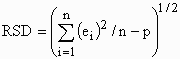 where ei is the residual value for the
ith observation, n is the number of observations, and p is the
degrees of freedom for the model. The RSD of the two exponential functions were
compared to the RSD of the linear-quadratic and allometric functions. Only
exponential equations with lower RSD than both simpler functions
(linear-quadratic and allometric) were considered. The marginal growth of each
component (lb) per pound of live weight gain was modeled as the derivative of
the function relative to live weight at each live weight.
where ei is the residual value for the
ith observation, n is the number of observations, and p is the
degrees of freedom for the model. The RSD of the two exponential functions were
compared to the RSD of the linear-quadratic and allometric functions. Only
exponential equations with lower RSD than both simpler functions
(linear-quadratic and allometric) were considered. The marginal growth of each
component (lb) per pound of live weight gain was modeled as the derivative of
the function relative to live weight at each live weight.
Results
The exponential function provided the best fit for total carcass fat, lipid-free soft tissue, and non-lipid fat tissue mass. The exponential nonlinear function provided the best fit for empty body protein and fat-free lean mass.
Empty body protein mass increased with live weight (Figure 1). As expected, the marginal growth of empty body protein decreases as live weight increases (Figure 2). The marginal growth of empty body protein is greater for gilts than barrows at all live weights.
Fat-free lean mass was greater for gilts than barrows, especially after 220 lbs live weight (Figure 3). The marginal growth of fat-free lean, which is the additional pounds of fat-free lean gain per pound of live weight gain at each live weight, is greater for gilts than barrows (Figure 4). However, the difference between barrows and gilts in marginal growth of fat-free total lean decreases as live weight increases and is very small after 240 lbs live weight. Overall, the marginal growth of fat-free lean has a consistent ratio to the marginal growth of empty body protein.
Total carcass fat mass increased more rapidly for barrows than gilts (Figure 5). The marginal growth of carcass fat tissue increases and starts to plateau at 290 lbs live weight (Figure 6). At 250 lbs live weight, approximately 36% of the gilts' and 48% of the barrows' live weight gain was carcass fat tissue. On a carcass weight basis, the marginal growth of carcass fat was approximately 45% for gilts and 60% for the barrows at 290 lbs live weight.
The percentage non-lipid components (water, protein, ash) in the incremental growth of the fat tissue is presented in Figure 7. The compositional growth of the fat tissue changes substantially as the pig grows. At 60 lbs live weight, approximately 44% of the fat tissue growth is non-lipid components. As the pigs grow, the incremental gain of the fat tissue is a higher percent lipid (76 to 78% at 250 lbs) and only 22 to 24% non-lipid components. For this reason, the energy cost (Mcal/lb) of carcass fat tissue growth increases as the pigs increase in weight.
Lipid-free soft tissue is a composite variable, as it includes fat-free lean mass and the non-lipid mass of the carcass fat tissue. Because lipid-free soft tissue growth is a complex variable, including both muscle (fat-free lean) and the non-lipid component of fat growth, it has a complex marginal growth rate (Figures 8 and 9). The growth of fat-free lean and carcass fat have drastically different nonlinear relationships with live weight. Also, the marginal compositional growth (% lipid) of the carcass fat tissue changes with live weight. For these reasons, lipid-free soft tissue mass has a multi-phasic relationship with live weight. The marginal growth of lipid-free soft tissue increases from 60 to 110 lbs live weight, as the marginal rate of fat-free lean gain is high and the marginal growth of carcass fat tissue, although low, contains a high percent non-lipid components. From 110 to 240 lbs, the marginal growth of lipid-free soft tissue decreases as the marginal growth of fat-free lean decreases and the compositional growth of the fat tissue, although increasing, contains a decreasing percent non-lipid components. After 270 lbs, the marginal growth of lipid-free soft tissue increases slightly as the rate of fat-tissue growth increases. The ratio of lipid-free soft tissue growth to either fat-free lean gain or empty body protein accretion is substantially different for different genetic populations, sexes and live weight body ranges. The ratio of lipid-free soft tissue to fat-free lean gain is {fat-free lean gain + [carcass fat tissue gain ´ (1 - % lipid in the fat tissue)]}/fat-free lean gain. Any factor (nutrition, genetics, environment, feed intake, or sex) affecting any of the three variables (fat-free lean gain, carcass fat gain, or % lipid in the fat tissue) would affect the ratio. The ratio of the marginal growth of lipid-free soft tissue mass to the marginal growth of fat-free lean or empty body protein is quite inconsistent and variable at different live weight ranges.
Implications
Fat-free lean gain is a measure of muscle growth that has consistent relationships with live weight and empty body protein mass. Lipid-free soft tissue growth is a complex variable, which has inconsistent relationships to empty body protein and muscle growth. The decrease in the marginal growth of muscle and increases in the marginal growth of carcass fat tissue and percent lipid within the fat tissue causes the decrease in the efficiency of both live weight and fat-free lean growth as live weight increases.
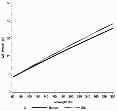
Figure 1. The relationship of empty body protein to live weight.
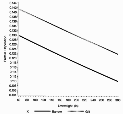
Figure 2. The marginal growth of empty body protein per pound of live weight gain.
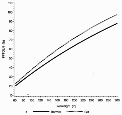
Figure 3. The relationship of fat-free lean to live weight.
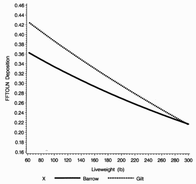
Figure 4. The marginal growth of fat-free lean per pound of live weight gain.
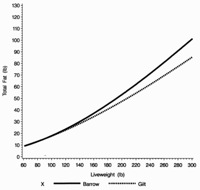
Figure 5. The relationship of total carcass fat mass to live weight.
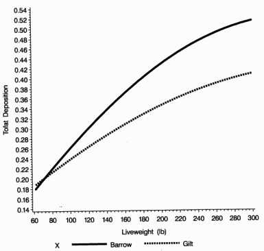
Figure 6. The marginal growth of carcass fat mass per pound of live weight gain.
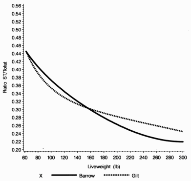
Figure 7. The marginal growth of non-lipid fat tissue per pound of total carcass fat gain.
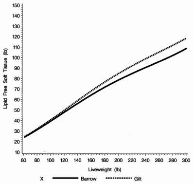
Figure 8. The relationship of lipid-free soft tissue mass to live weight.
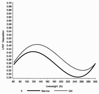
Figure 9. The marginal growth of lipid-free soft tissue per pound of live weight gain.